

Atlas – C1 Vertebra
Yes, Atlas. The Greek titan who held up the celestial spheres.
Wait, we’re talking about medicine, right. Oh well, guess I’ll have to talk about the significantly less cool (although the significantly more real) Atlas of our body, the C1 vertebra. And while we’re at it, let us also complete learning about all the vertebrae in the body, and finish the Axis, C2, as well.
Remember, for a review of the Cervical Vertebrae, you can check out my blog post: https://theartofmed.wordpress.com/2015/05/23/what-is-this-blog-about/
Back to the Atlas and Axis, these two vertebrae are together called the craniovertebral vertebrae, due to their very important articulations with the skull, as will be discussed lower down.
Let us begin with the Atlas for now.
Atlas
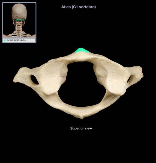
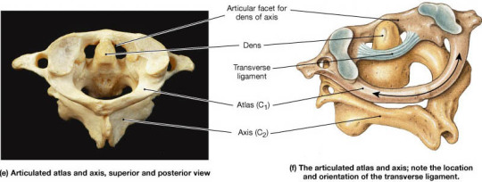
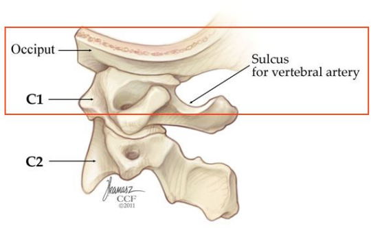
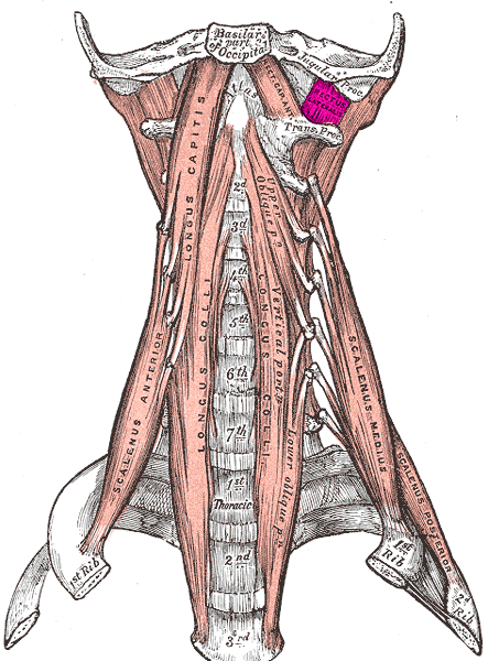
The atlas is the 1st Cervical vertebra. It directly articulates with the base of the skull, as shown in this figure below:
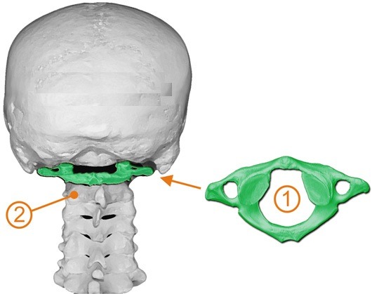
Here is the Atlas, colored in green.
What is the most striking thing you notice about it when you compare it to other vertebrae below?
I’ll tell you what I see, I see that the Atlas is somehow much wider than the other vertebrae. It’s as if the C1 vertebra is lifting its hands and holding up the skull, our own celestial sphere, or the globe of our head. It really is like Atlas, isn’t it? Holding out its “hands” and holding the weight of the skull up, just as Atlas Himself did.
It is obvious it is an irregular vertebra, because it’s function is definitely different from the other Cervical Vertebrae.
Structure of Atlas
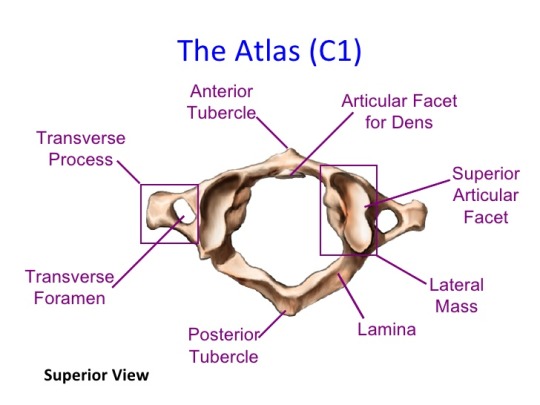
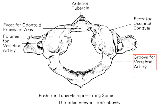
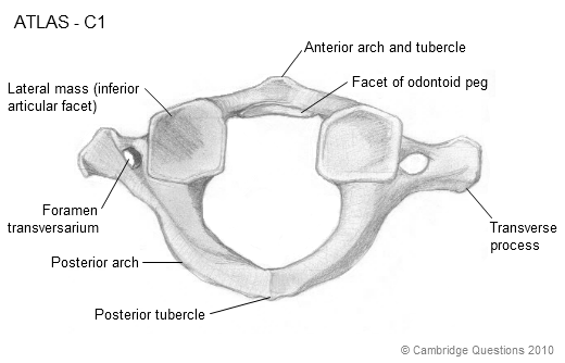
The atlas is irregular because it contains no body nor spinous process, and is much wider than the other cervical vertebrae.
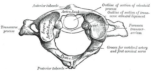
It can thus be described as a ring-shaped vertebra, with the ring divided into an anterior arch in the front and a posterior arch in the back, each separated from one another by laterally extending structures collectively called the lateral masses, and transverse processes.
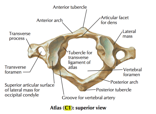
Thus, there are 4 main features we need to consider about the Atlas:
- Short Anterior Arch
- Long Posterior Arch
- Lateral Masses
- Transverse Processes
Anterior Arch
The anterior arch forms about 1/5 of the ring. The anterior surface is convex and projects outwards, in an anterior direction. It serves as a rudimentary body of the C1 vertebra.
The most anterior part of the anterior arch forms the anterior tubercle.
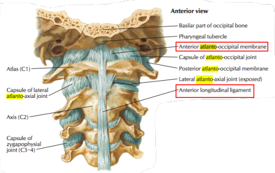
This anterior tubercle, shown above in green, is extremely important for the attachment of a ligament, the anterior longitudinal ligament.
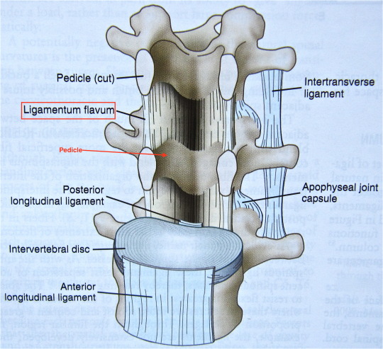
The anterior longitudinal ligament is a strong, fibrous band that runs along the anterolateral aspects of vertebrae and IV discs, extending superiorly from the pelvic (anterior surface) surface of the sacrum to the anterior tubercle of the Atlas. The anterior longitudinal ligament is thin over the vertebrae, but thick over the IV discs, and functions as the only ligament to prevent hyperextension of the vertebral column. The anterior longitudinal ligament also extends superiorly above the anterior tubercle, to connect to the anterior surface of the foramen magnum as the anterior antlanto-occipital membrane, a membrane attached to the sueprior border of the anterior arch. Furthermore, the region of the anterior longitudinal ligament that connects the atlas to the axis is the anterior atlanto-axial ligament, a membrane attached to the inferior border of the anterior arch. Both of these will be explained later.
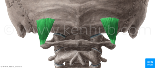
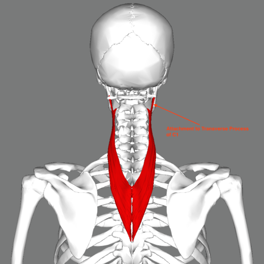
On either side of the midline, the anterior tubercle also gives attachment to the superior oblique part of the longus colli muscle. This it the whole muscle here:
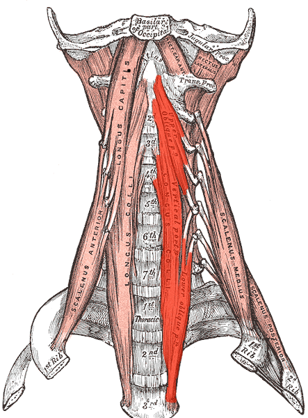
The longus colli muscle is greek for long muscle of the neck, and we can see why: The longus colli muscle spans superiorly from the anterior tubercle of the atlas, as stated before, inferiorly until the T3 vertebra.
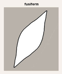
Looking at the diagram, it appears to be somewhat fusiform shaped, i.e, wide in the middle, and pointed on either end, somewhat like this:
With this in mind, the longus colli muscle is divided into 3 parts:

1) Superior Oblique Portion: The superior oblique portion originates from the anterior tubercles of the transverse processes (remember the anterior tubercles of the normal vertebrae are located on the transverse process. The anterior tubercle of the Atlas is in the midline) of C3, C4 and C5 vertebrae. It ascends obliquely (hence superior oblique) and in a medial direction, and attaches to the anterior tubercle of the atlas.
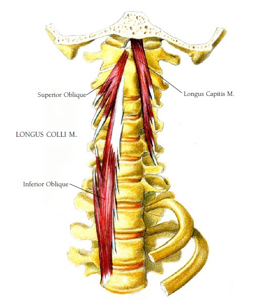
2) Inferior Oblique Portion: The inferior oblique portion is the smallest part of the longus colli muscle. It originates from the front of the bodies of T1, T2 and sometimes T3. You can visualize this in the diagram above. It ascends obliquely and laterally inserts to the anterior tubercles of the transverse processes of C5 and C6.
3) Vertical Portion: The vertical portion literally ascends vertically, originating from the anterior part of the bodies of C3/C4 – C5 and T1-T3. After ascending vertically, it inserts into the anterior portion of the bodies of the C2-C4 vertebrae.
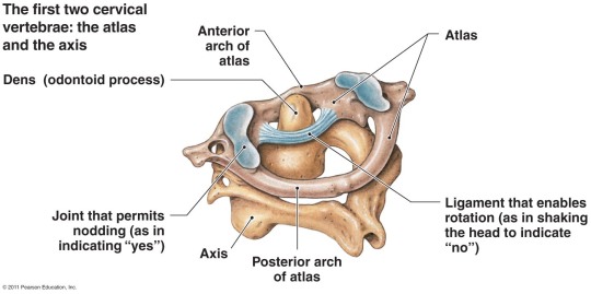
On the other hand, the posterior surface of the anterior tubercle holds an oval shaped facet. This facet attaches a structure known as the odontoid process/dens of the axis, a long tooth-like projection that extends from the C2 vertebra.
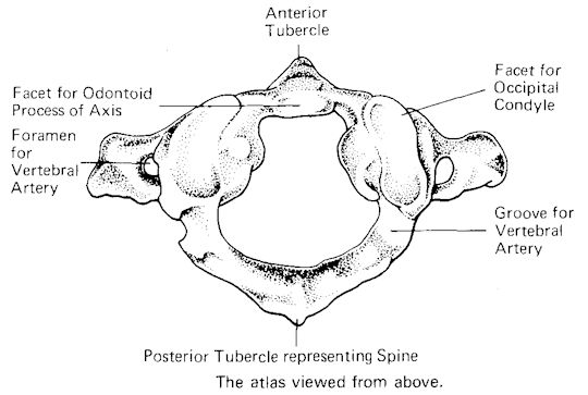
Notice where the label is in the diagram above.
Now look at how the odontoid process looks below:
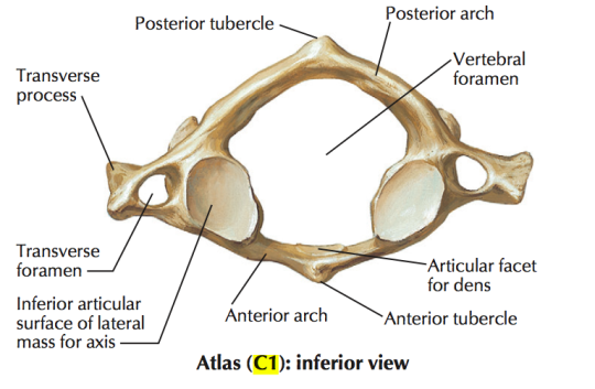
Posterior Arch
The posterior arch forms about 2/5 of the ring structure. It extends posteriorly until the posteriormost tubercle, the posterior tubercle of the atlas. The posterior tubercle serves as a rudimentary spinous process of the C1, since no proper spinous process exists.
Posterior Tubercle of Atlas:
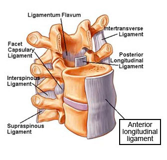
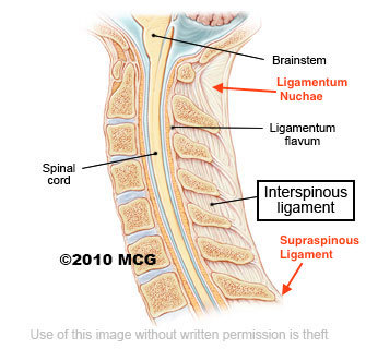
Looking at the diagram above, you can see the posterior arch is larger, and ends in a posterior tubercle as its most posterior region, in the midline. The posterior tubercle gives attachment to a ligament, known as the ligamentum nuchae.
The ligamentum nuchae is a superior continuation of the supraspinous ligament.
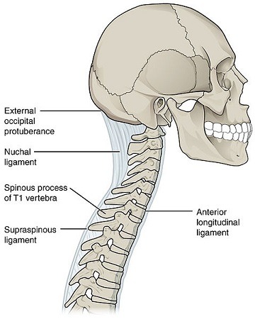
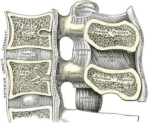
The supraspinous ligament is a thick, cord like fibrous band that connects the apices of the spinous processes, from C7 to the sacrum. It is continuous with the ligamentum nuchae above C7, and it continuous with interspinous ligaments between the spinous processes. The interspinous ligaments and thin, fibrous membranes that connect adjacent spinous processes together, by running obliquely from the root of the spinous process to the apex. They are continuous with the supraspinous ligament posteriorly, and continuous with a ligament known as the ligamentum flavum anteriorly (we discuss this ligament below). The interspinous ligaments are narrow in the thoracic region, but thicker in the lumbar region. In the cervical region, they are very underdeveloped, and are considered part of the ligamentum nuchae. We will see why just now.
Both the supraspinous ligament and the interspinous ligament prevent hyperflexion of the vertebral column.
Now, it is important to remember that C7 is the vertebra prominens, with the largest spinous process of the body. Beyond this point, the supraspinous ligament is not able to change its direction so as to directly come into contact with the spinous processes of vertebrae C6 and above. Hence, instead, the supraspinous ligament gives off extensions that spread to the spinous processes of the C1- C6 vertebrae and this forms a median, fibro-elastic band known as the ligamentum nuchae.
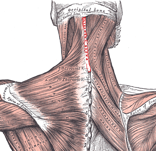
The ligamentum nuchae extends superiorly up till the external occipital protuberance of the skull, visible in the diagrams above. The important thing about this ligament is its location. Because it is located in the midline, and it is located over the very short and deeply located spinous processes of the C1-C6 vertebrae, it:
Compartmentalizes the neck into a left and right compartment.
Allows attachment for muscles inserting into the midline above the C7 spinous processes. These muscles are the splenius capitis and upper fibres of the trapezius.
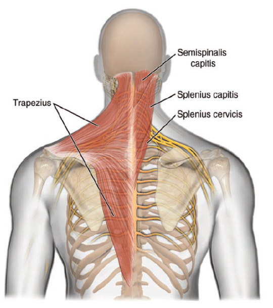
[Note that although the semispinalis captious and splines cervicis are labelled here, the semispinalis capitis is inserted deep to the ligamentum nuchae, and splenius cervicis is inserted below the ligamentum nuchae.]
On either side of the posterior tubercle is the insertion of the rectus capitis posterior minor muscle.
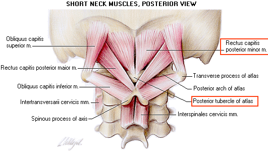
Furthermore, the inferior surface of the posterior tubercle attaches a very small, deep muscle of the back, the interspinalis cervicis.
Don’t worry about all the muscles you’re seeing here, when we cover muscles of the neck, we’ll go through these in great detail.
Superior Surface of Posterior Arch:
The superior surface of the posterior arch contains a groove for the vertebral artery and C1 spinal nerve (suboccipital nerve). This groove is located just adjacent to the lateral masses of the Atlas.
Directly behind this groove, the superior surface of the posterior arch provides attachment to the posterior atlanto-occipital membrane.
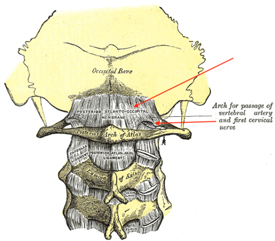
The posterior atlanto-occipital membrane is a broad, thin fibrous membrane that extends from the superior surface of the posterior arch, superiorly till the posterior margin of the foramen magnum.
As seen in the diagram above, there is a defect in the inferolateral part of the membrane that allows the passage of the C1 spinal nerve and vertebral artery.
Inferior Surface of the Posterior Arch:
The inferior surface of the posterior arch provides attachment for the posterior atlantoaxial membrane. It is a broad, thin ligament that attaches superiorly to the inferior surface of the posterior arch, and inferiorly to the superior surface of the lamina of the axis, the C2 vertebra.
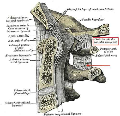
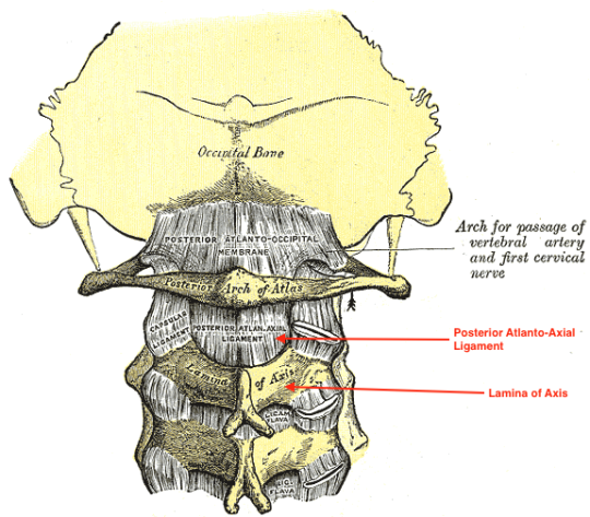
Both the posterior atlanto-occipital membrane and posterior atlanto-axial ligament are continuations of the ligamentum flavum. The ligamentum flavum is a yellow, elastic ligament that extends between the pedicles of the vertebrae. It begins inferiorly at the 1st Sacral segment below, and extends up till the Body of the C2 Vertebra, the Axis. Above the C2 vertebra, it forms the posterior atlanto-axial membrane till the Atlas, and the posterior atlanto-occipital membrane from the Atlas to the Occipital Bone.
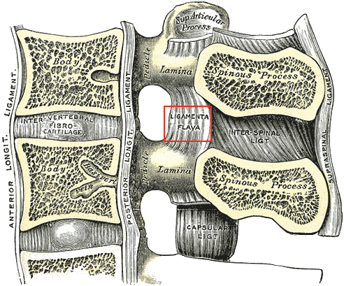
These ligaments extend almost vertically from one lamina to another, and they meet and blend with the ligamentum flavum on the opposite side along the midline. They are thinnest in the cervical region, but get thicker in the thoracic region, and thickest in the lumbar region. The marked elasticity of the ligamentum flavum keeps the vertebral column rigid and fixed in place, preventing abrupt flexions of the vertebral column that may damage it.
Lateral Masses
The lateral masses are the most bulky part of the Atlas, very large (like the muscles of Atlas) in order to support the weight of the globe of the head, which is our own celestial sphere.
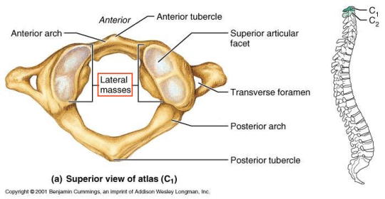
The superior view, seen above, shows the lateral mass, labelled. The superior part of the lateral mass shows a kidney shaped, concave superior articular facet, that articulates with the occipital condyles of the base of the skull, to form the atlanto-occipital joint.
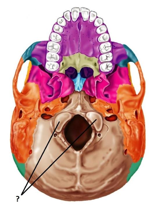
The “ ? “ above shows the occipital condyles, also kidney shaped, but convex or protruding outwards. Notice then that it shows a perfect complement to the Atlas. No wonder our head sits so well, Atlas is doing a great job.
This atlanto-occipital joint is formed between the occipital condyles and the superior articular facet of the lateral mass of the Atlas. The joint is a condyloid type synovial joint. Look at how the joint is shaped:
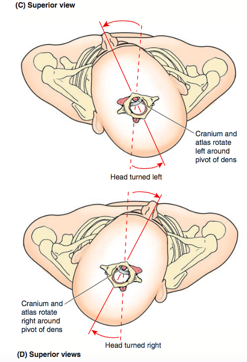
The joint functions like a seesaw:
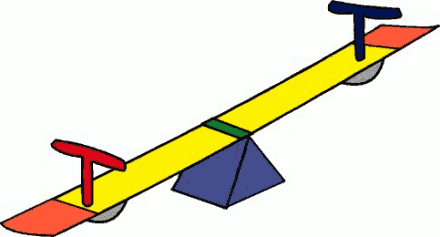
Imagine each end of the seesaw is the direction in which you move your head. The little pivot, or triangle in the middle in this case is the Atlas, and it will allow the seesaw to move towards the direction more weight is applied in. Thus, if you want to say yes, and move your head forwards then backwards, it is the atlanto-occipital joint that allows this movement, by moving the occipital condyles within the superior articular facet of the lateral mass. We can thus say the atlanto-axial joints allow a ‘yes’ motion (extension and flexion of the neck) and also allows a small amount of lateral flexion (sideways tilting, as in confusion).
Medial Surface of Lateral Mass:
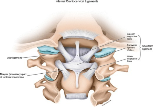
The lateral mass contains a small tubercle for the attachment of a ligament known as the transverse ligament of the atlas, that extends from the medial surface of one lateral mass to the medial surface of the lateral mass on the opposite side.
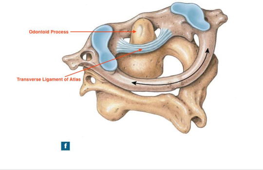
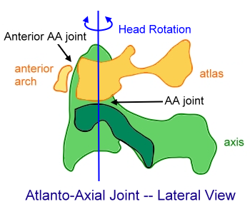
Notice that the transverse ligament of the atlas bounds the odontoid process posteriorly. This is known as the median atlanto-axial joint, formed between the odontoid process, the posterior surface of the anterior tubercle, and the transverse ligament of the atlas. This is a pivot type synovial joint. The black arrows in the diagram very effectively illustrate what kind of movement we can expect from the median atlanto-axial membrane.
To picture this, imagine Emma Stone playing with a hula hoop. The hula hoop swings around Emma’s body in circles as she play with it.

Superimposing that onto the median atlanto-axial joint, the odontoid process or dens is Emma Stone, and the entire Atlas is the hula hoop, able to swing all the way around the odontoid process! But thankfully it doesn’t. It is restricted by a joint described immediately below, that restricts the degree of rotation allowed by the median atlanto-axial joint. Thus, the main motion allowed by the media atlanto-axial joint is rotation, allowing us to say “no” with our heads. The transverse ligament of the atlas is very important so as to keep the odontoid process in place.
Inferior Surface of Lateral Mass:
The inferior surface of the lateral mass contains the inferior articular facet. This inferior articular facet is nearly circular, is more or less flat, and articulates with the superior articular facets of the Axis, to form the lateral atlanto-axial joint.
Notice how round the inferior articular facet looks from the inferior surface. It forms 2 lateral atlanto-axial joints, and these joints are gliding synovial joints, that function mainly to restrict the degree of motion of the median atlanto-occipital joint.
There are also a number of ligaments associated with the atlanto-axial joints that restrict the degree of movement of the odontoid process and atlanto-axial joints. These ligaments are the apical ligament, alar ligament, and cruciate ligament.
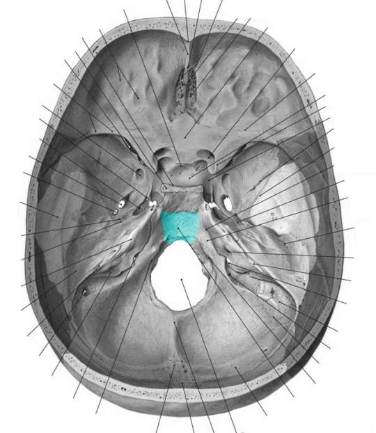
The apical ligament is a thin, string like ligament that extends from the very apex of the odontoid process and attaches just onto the clivus of the occipital bone, passing through the foramen magnum. The clivus is a region located on the inside of the skull, on the basilar part of the occipital bone. It is shown in blue below, ignoring all the lines:
It prevents the odontoid process from moving too inferiorly in relation to the Atlas.
The alar ligaments are a pair of cord like ligaments that extend from each side of the odontoid process, and attaches to the medial ends of the occipital condyles. It prevents lateral displacement or sideways displacement of the odontoid process.
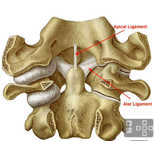
The cruciate ligament is actually a collection of 3 ligaments. These are the transverse ligament of the atlas, superior longitudinal band, and inferior longitudinal band. We have discussed the transverse ligament of the atlas already. The superior and inferior longitudinal bands are simply superior and inferior extensions from the middle of the transverse ligament of the atlas. This results in a cross-shaped structure of ligaments, and hence the name, cruciate (cross like). The superior longitudinal band extends superiorly from the transverse ligament of the atlas, attaching on the clivus deeper through the foramen magnum than the apical ligament. The inferior longitudinal ligament extends inferiorly from the inferior border of the transverse ligament of the atlas towards the body of the axis, where it attaches.
[The superior longitudinal band is blocking the apical ligament from view].
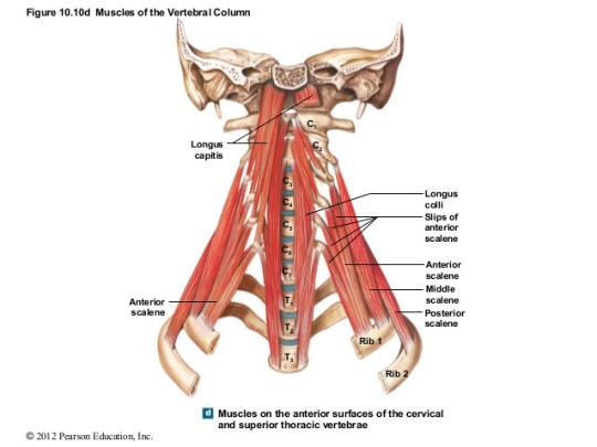
The anterior surface of the lateral mass also attaches a muscle, the rectus capitis anterior muscle. This muscle is a short, flat muscle of the anterior neck situated immediately behind the longus capitis muscle. From its origin on the anterior surface of the lateral mass of the atlas, it ascends obliquely in a medial direction to insert in the inferior surface of the basilar part of the occipital bone, on the anterior margin of the foramen magnum. This muscle actually is the muscle that helps you look down! So it is a flexor of the head.
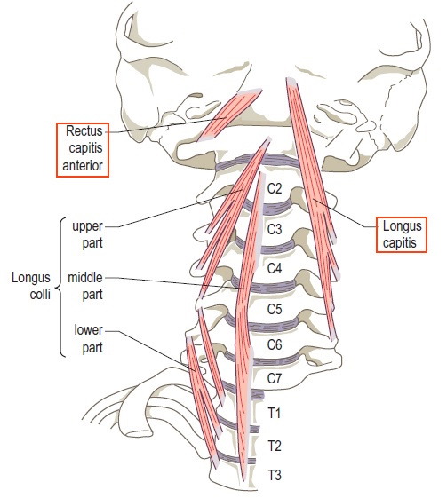
Note in the picture above, that the longus capitis muscle is cut off on the left side, and hence does not block the rectus capitis anterior anymore.
Transverse Processes
The transverse processes are the lateralmost structures of the Atlas, extending laterally from the C1 vertebra’s lateral masses. It is actually quite long, and can be felt between the angle of the mandible and mastoid process. It indeed, contains features typical and expected of the transverse processes of the regular cervical vertebrae. This means that the transverse process still contains the most characteristic feature, the foramen transversarium, that transmits the vertebral artery.
Recall that there is also a groove on the superior surface of the posterior arch just adjacent to the lateral masses. Thus, we can see how the vertebral artery travels along the C1 vertebra: It travels through the foramen transversarium and then immediately enters the groove for the vertebral artery on the superior surface of the posterior arch. From here, it travels through the defect in the posterior atlanto-occipital membrane.
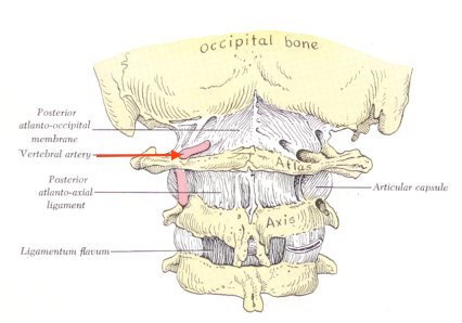
Notice it travels through the foramen transversarium, then over the groove, then straight through the membrane in the picture above.
It also provides attachment to a large number of muscles.
These muscles include:
1) Rectus Capitis Lateralis: Has origin from superior surface, anteriorly, of transverse process of C1, and inserts into the jugular process of occipital bone.
2) Obliqus Capitis Superior Muscle: Has origin at superior surface, posteriorly of transverse process of C1, and inserts into the lateral half of the inferior nuchal line.
3) Obliqus capitis Inferioris Muscle: Originates from the apex of the spinous process of the C2 vertebra (Axis) and inserts into the posterior surface of the transverse process of C1, close to the tip.
4) Levator Scapulae Muscle: This muscle originates from the posterior tubercles of the transverse processes of C2-C4 and the lateral margin and lower border of the transverse process of C1, the atlas. It inserts into the upper part of the medial border of the scapula, and the superior angle, as discussed under “Scapula.”
5) Splenius Cervicis: This muscle originates from the spinous processes of T3-T6, and inserts into the posterior tubercles of C2 and sometimes C3, and the transverse process of C1.
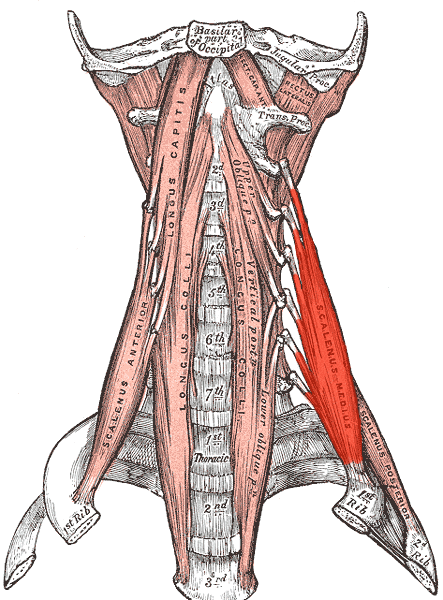
6) Scalenus Medius: This muscle originates from the transverse process of C1-C6, and inserts into the upper surface of the 1st rib.
7) There are an additional two deep instrinsic muscles of the back, the intertransversarius posterior cervicis and intertransversarius anterior cervicis, attached to the transverse process.
Occasionally, the transverse process sometimes fuses with the occipital bone, a process known as occipitalization, and this is a serious condition that can compress the spinal cord.
Vertebral Canal
We already know the vertebral canals of cervical vertebrae are large. However,the vertebral canal of the C1 vertebra is divided into two compartments by the transverse ligament of the atlas. As visualized above, the odontoid process passes through the anterior compartment. It is the spinal cord that passes through the posterior compartment.
Thus, if the transverse ligament of the atlas is ever torn, the odontoid process may compress the spinal cord causing death.
Ossification of Atlas
The atlas is ossified from 3 centres, by endochondral ossification.
One appears in each lateral mass by the age of around 7 weeks, and extends posteriorly to form the posterior arches. By the age of 3-4, they fuse in the midline to form a posterior tubercle either by fusion, or by the use of a cartilage medium formed by a separate centre.
At birth, the anterior arch consists of only cartilage, but produces an ossification centre, which converts the anterior arch to bone.
That’s all guys! Hope this post helped you all!
VIDEOS:

Thank you! Really helped me connect bone and muscle anatomy
LikeLike
Wonderful explanation!
LikeLike