

Inclusion bodies are those things you look at in cells and think, “Why are you even here?” Inclusion bodies are those things that just appear in cells and.. don’t really do anything. They’re just included within the cell, for some purpose.
Typically, inclusion bodies are nuclear or cytoplasmic aggregates of stainable substance, usually proteins.
The inclusion bodies in red blood cells are almost always indicative of some sort of pathology, and thus it is useful to understand each inclusion body that can occur within a red blood cell.
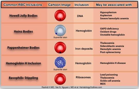
This diagram is a pretty comprehensive summary of most of (not all) the inclusion bodies we will come across. But as usual, let us go through each in detail:
Inclusion Bodies Within Red Blood Cells:
- Howell-Jolly Body
- Heinz Body
- Hemoglobin H Inclusion
- Pappenhemier Body
- Basophilic Stippling
- Cabot Ring
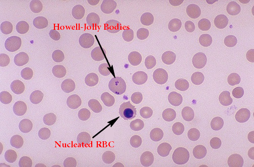
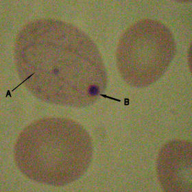
Howell-Jolly Body
Howell-Jolly bodies are histopathological findings on a blood smear. They appear as a very dark purple (described as a basophilic area) spot within the cytoplasm of the red blood cell. They are in fact, basophilic nuclear remnants, or remnants of DNA.
Typically, they are round to oval shaped, 1um across and are usually restricted to only 1 per cell, although there can be multiple amounts.
Typically, during the maturation of a red blood cell in the bone marrow, the nucleus must be expelled alongside all nuclear matter in order to become a mature red blood cell. However, in rare occasions, the nucleus is not appropriately removed and fragments of the DNA remain as these basophilic dark spots, and are called Howell-Jolly bodies. They are usually produced physiologically, but are quickly and easily removed by the spleen, that identifies the Howell-Jolly body and identifies the cell as defective.
Then, if Howell-Jolly bodies are present in high amounts, it must indicate that something is wrong with the spleen. If the spleen is malfunctioning in some way, then Howell-Jolly bodies are not removed and they remain in circulation.
Diseases Associated with Howell-Jolly Bodies:
- Splenectomy
- A splenectomy would leave an individual with no spleen to remove malformed red blood cells. Thus, even if red blood cells have Howell-Jolly bodies within their cytoplasm, the absence of a spleen means that these inclusion bodies will not be removed and are readily seen on a blood film.
- Hyposplenism
- Same as aforementioned explanation, except that the spleen is poorly functional and does not remove Howell-Jolly bodies readily.
- Hyposplenism can occur either from direct trauma, or via autosplenectomy, where a disease such as sickle cell anemia damage the spleen to such an extent that it becomes non-functional.
- Severe Megaloblastic Anemia
- Notice the word severe. In severe megaloblastic anemia, it is possible for the buildup of chromosomal remnants as RNA synthesis continues unhindered. The mechanism for this is still under investigation however.
- Severe Hemolytic Anemia
- As blood loss occurs severely, the bone marrow is pressured to produce more red blood cells. Thus, there is a “left-shift” towards more erthyrcoytes being produced, even if they are immature. It is thus possible for red blood cells with whole nuclei being produced in this condition. Often however, the nuclei are still broken down into Howell-Jolly bodies, but are produced in such sheer numbers due to the left shift that the spleen cannot remove all of them.
Note that it is also possible for red blood cells to be entirely nucleated. A nucleated red blood cell, or NRBC, is a red blood cell (RBC) that retains nucleus. Normally, NRBCs are only found in the circulation of fetuses and newborn infants. After infancy, RBCs normally only contain an nucleus during the very early stages of the cell’s life, and the nucleus is ejected before the cell is released into the bloodstream. Thus, if NRBCs are seen on an adult’s peripheral blood smear, it suggests that there is a very high demand for the bone marrow to produce RBCs, and immature RBCs are being released into circulation. Possible pathologic causes include anemia, myelofibrosis, thalassemia, miliary tuberculosis, cancers involving bone marrow, and in chronic hypoxemia.

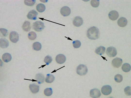
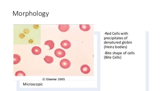
Heinz Body
Do you remember what “Bite cells” are from “Morphological Abnormalities of Red Blood Cells”? They are also referred to as Degmacytes, and occur when a portion of a red blood cell is phagocytosed due to the presence of an inclusion body within the cytoplasm of the cell. This inclusion body is the Heinz Body.
Heinz bodies are formed by damage and denaturing to the hemoglobin component of red blood cells, most commonly by oxidative stress, but also possibly by genetic abnormalities in hemoglobin.
Typically, during oxidative damage to hemoglobin, an electron is transferred from the hemoglobin to oxygen, resulting in the formation of a reactive oxygen species (ROS). This ROS can lead to severe damage within the cells, and can even cause lysis of the entire cell.
The ROS denatures portions of the hemoglobin, causing the to precipitate and produce Heinz Bodies, which becomes an antigenic agent. Thus, macrophages detect the antigen and remove the damaged portions of the cell, its damaged membrane and the denatured hemoglobin (now called the Heinz Body).
Diseases Associated with Heinz Bodies:
Heinz bodies are almost always associated with oxidative damage to the red blood cell, and only rarely to genetic causes. So obviously then, the cause of Heinz Bodies would be something that causes increased oxidative stress to increase the likelihood of production of ROS in the blood cells. So what are these conditions?
- Glucose-6-phosphate dehydrogenase Deficiency (G6PD Deficiency)
- G6PD deficiency is the most important and most common cause of production of Heinz Bodies, and consequently Degmacytes.
- In G6PD deficiency, the loss of the G6PD enzyme has serious consequences that increase oxidative stress.
- Normally, G6PD as an enzyme is used in the pentose phosphate (or HMP Shunt) pathway.
- G6PD converts glucose-6-phosphate into 6-phosphoglucono-δ-lactone and is the rate-limiting enzyme of this metabolic pathway that supplies reducing energy to cells by maintaining the level of the reduced form of the co-enzyme nicotinamide adenine dinucleotide phosphate (NADPH).
- The NADPH in turn maintains the supply of reduced glutathione in the cells that is used to mop up free radicals that cause oxidative damage.
- The G6PD / NADPH pathway is the only source of reduced glutathione in red blood cells (erythrocytes). The role of red cells as oxygen carriers puts them at substantial risk of damage from oxidizing free radicals except for the protective effect of G6PD/NADPH/glutathione.
- NADPH Deficiency
- NADPH deficiency can cause a deficiency in glutathione peroxidase, which is an enzyme that can reduce hydrogen peroxide, a ROS, into water.
- Chronic Liver Disease
- Alpha Thalassemia
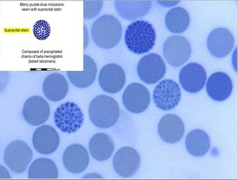
- Normal adult hemoglobin is composed of two alpha and two beta chains. Alpha thalassemia patients have partial or complete defects in alpha globin production, leading to a relative abundance of beta globin chains in the cell. These excess beta globin chains aggregate to form HbH, which has decreased solubility and precipitates in the red blood cell cytoplasm. This is not direct damage to hemoglobin per se, but rather a perturbation in the quaternary structure of hemoglobin.
- Thus, HbH is merely a moderate to severe form of alpha-thalassemia that consists of hemoglobin precipitating within the cytoplasm of the red blood cells in high amounts due to the insoluble nature of beta-chain heavy hemoglobin molecules.
- Because the HbH aggregates are precipitated and are non-functional, HbH is considered a subtype of Heinz Bodies.
Observe a HbH aggregate below:
- *Hyposplenism and Splenectomy
- While not a direct cause of Heinz Bodies, the spleen helps with clearing the blood and thus removing Heinz Bodies from circulation. With a damaged spleen, more of these Heinz Bodies remain in circulation and are visible in a blood slide.
Observe a Heinz Body below:
Difference Between Heinz Body and Howell-Jolly Body:
Howell-Jolly bodies can only be seen with a special supravital stain (a supravital stain is a staining technique that involves using a stain on cells that are removed from the body), like the new methylene blue stain. On the other hand, the Howell-Jolly bodies are seen as very dark, basophilic spots on the cell.
Furthermore, the Heinz Bodies are usually found close to the inner surface of the RBC membrane, while Howell-Jolly bodies can be found in variable positions around the cell.
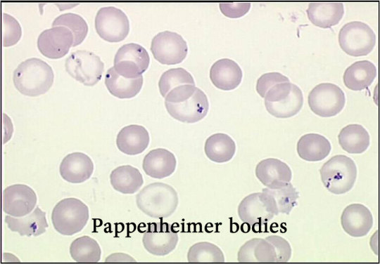
Pappenheimer Body
Pappenheimer bodies come next. We discussed hemoglobin deposits (HbH and Heinz Bodies) and nuclear inclusion bodies (Howell-Jolly Bodies), but the Pappenheimer Bodies are inclusion bodies of iron. They are deposits or iron that build up within the red blood cell.
Pappenheimer bodies are basophilic erythrocytic inclusions that are usually located at the periphery of the cell. They contain iron and stain with Prussian blue. Prussian blue is the stain that is used to identify that these Pappenheimer bodies are pure iron deposits, and not heme as in Heinz Bodies.
Cells containing Pappenheimer bodies can be confused with late reticulocytes. Prussian blue stain, which is not taken up by reticulocytes, is helpful in differentiating the two. Pappenheimer bodies can also cause a false elevation of platelet counts when performed with electronic counters. Pappenheimer bodies are also visible with a Wright and/or Giemsa stain, but Prussian blue is more useful to differentiate these bodies from Heinz Bodies.
Diseases Associated with Pappenheimer Bodies:
- Splenectomy
- Due to inability of spleen to remove pappenheimer laced cells.
- Hemolytic Anemia
- Sideroblastic Anemia
- Megaloblastic Anemia
- Hemoglobinopathies
The exact cause of Pappenheimer bodies in these diseases is unknown.
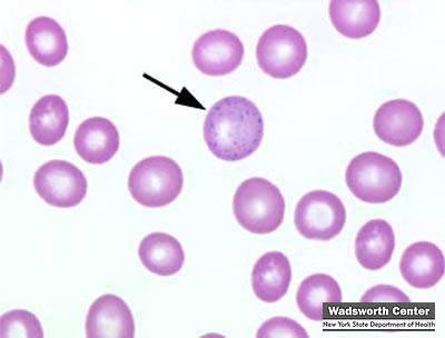
Basophilic Stippling
Basophilic stippling is also referred to as punctuate basophilia. In this condition, there are fine and coarse granules located at in a fairly evenly distributed manner . These small dots are actually accumulations of RNA and ribosomes.
Diseases Associated With Basophilic Stippling:
Basophilic Stippling can be divided into fine basophilic stippling and coarse basophilic stippling.
- Fine basophilic stippling is associated with increased red cell production and is commonly seen when there is increased polychromatophilia.
- Coarse basophilic stippling is seen in megaloblastic anemia and other forms of severe anemias, lead poisoning, and thalassemia. Coarse basophilic stippling indicates impaired hemoglobin synthesis, probably due to the instability of RNA in the young cell.
In fact, basophilic stippling is usually a very strong sign of lead poisoning.
Cabot Ring
Cabot rings. These things are really interesting. They are literally loops, rings or figure 8 type structures that are located within the cytoplasm of the RBC. They may look like beads on a string. They are typically colored red-purple under the Wright Stain.
They are quite rare, but they look really cool whenever you come across one. They are simply microtubular remnants of mitotic tubules that are involved in mitosis.
Their presence usually is indicative of some abnormality in the production of red blood cells.
Diseases Associated with Cabot Rings:
- Myelodysplastic Syndrome
- Megaloblastic Anemia
Both these conditions show abnormalities in production of red blood cells.

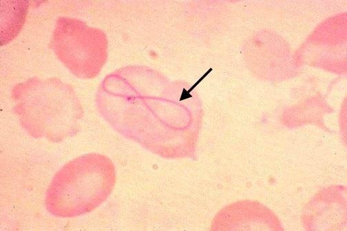
And here is a very unlucky cell with both Cabot rings and a Heinz Body:
That’s it for all the abnormalities guys! Hope this was useful as usual! You can combine this with “Morphological Abnormalities of RBCs” to figure out what pretty much anything strange about an individual red blood cell indicates. As usual, follow for more content!

Excellent presentation. Great help for professors and students.
LikeLike
absolutely stunning presentation .. I solute u for this …God bless you
LikeLike
excellent presentation ..you are a great teacher
LikeLike
Perfect way of high class education
LikeLike
Great! Thanks for the explanations!
LikeLike
This was very useful thanks a lot .
LikeLike
I am a veterinary clinical pathologist I found a cabot ring in a macro plate in a dog, can you find them in the platelets in humans? does it have any meaning according to you?
LikeLike
Good for reference, especially for medical lab scientists.
LikeLike
Superb
LikeLike
Thanks for the explanations ,has helped me alot
LikeLike
Correction: “Howell-Jolly bodies can only be seen with a special supravital stain” should read “Heinz bodies…”
LikeLike
In the following section, “Howell-Jolly” should actually be “Heinz.” Just thought you might want to edit to avoid confusion!
“Howell-Jolly bodies can only be seen with a special supravital stain (a supravital stain is a staining technique that involves using a stain on cells that are removed from the body), like the new methylene blue stain.”
Otherwise, awesome summary! Thanks.
LikeLike
Hi, thanks for the great info! I think there is a mix up in one of the sections; when you were talking about the difference between Heinz bodies and Howell-Jolly bodies. A supravital stain is needed to see the presence of Heinz bodies, not Howell-Jolly bodies.
LikeLike
This site is outstanding! Permit me to suggest one small correction. Where you state, “Howell-Jolly bodies can only be seen with a special supravital stain …”, I believe you should state “Heinz bodies” instead.
LikeLike
“Howell-Jolly bodies can only be seen with a special supravital stain (a supravital stain is a staining technique that involves using a stain on cells that are removed from the body), like the new methylene blue stain. On the other hand, the Howell-Jolly bodies are seen as very dark, basophilic spots on the cell.”
It should read– Heinz bodies can only be seen with a special supravial stain…
LikeLike
Well put about inclusion topic
LikeLike
great explanation , easy way to understand. Thank you
LikeLike
Thanks its very useful
LikeLike